Before we get into the science of fetal brain development heres a quick anatomy primer. During the second trimester your babys brain is directing the diaphragm and chest muscles to.
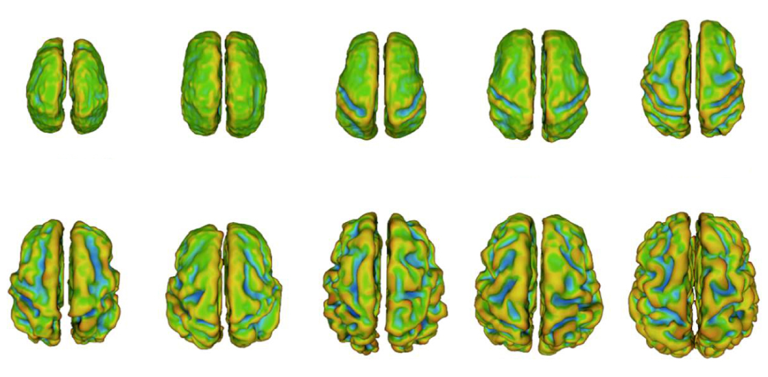 Brain Imaging Studies Seek Signs Of Autism Before Birth Spectrum Autism Research News
Specifically we first investigated the relationship between MRI-based linear biometric data and brain development in 637 normal fetuses in a wide range of gestation age from 22 to 40 weeks which were screened from a large local database.

Brain development in fetuses. Fetuses and infants to those with a high likelihood for developing autism defined by having an autistic mother or autistic sibling. From the eighth week until the moment of birth it is known as fetus. The bulk of cortical neurogenesis occurs during the mouse embryonic period corresponding to early fetal life in the human but is ongoing into postnatal life in the hippocampus and cerebellum.
The mechanisms underlying impaired fetal brain development in the setting of maternal stress are complex and multifactorial. Finally we aimed to establish an MRI-based. Prenatal imaging for diagnosis and prediction of neurodevelopmental outcomes in fetuses with brain malformations is a substantial challenge.
Did You Know. To our knowledge this is the first report indicating that maternal psychological distress is associated with impaired brain development in fetuses with CHD. Babys Nervous System and Brain The parts of your babys brain.
Brain development involves the formation of the brain nervous system and spinal cord it all begins at the embryonic stage itself. A mere 16 days after conception your fetuss neural plate forms think of it as. Cerebrum cerebellum brainstem and intracranial cavity.
The development of the brain in the fetus includes the development of the brain together with the nervous system and the spinal cord. Third-trimester brain volumes were reduced and brain growth trajectories were slower in the ex utero preterm group compared with the in utero healthy fetuses. Fetal brain development timeline In the first two weeks of pregnancy the fertilized egg is implanting inside of the uterus.
Accurate diagnosis of brain abnormalities has important therapeutic implications. Its the largest part of the brain and contains the. In the third week the heart the backbone and the brain of the embryo start to form.
Brain Development in the Fetus-The brain development stages start from the first week and go up to the 40th week-Brain Development Issues. Fetuses with CHD especially those with lowest cerebral substrate delivery show a regionspecific pattern of small brain volumes and impaired brain growth before 32weeks gestation. During fetal life the predominant source of brain energy is glucose which crosses the placenta by facilitated diffusion.
When Does the Fetuss Brain Begin to Work. How Your Babys Brain Develops First Trimester. Your babys brain will grow five main parts each responsible for a different aspect of directing the body and eventually the mind and decision making.
Genes contributes to about 60 of brain development environment in uterus to about 30 and maternal nutrition to about 10. Comparison with human development can be challenging as mice are postnatal brain developers with a gestational length of 19 to 21 days. The cerebrum is responsible for thinking feeling and memory.
Generally speaking the central nervous system which is composed of the brain and the spinal cord matures in a sequence from tail to head. The cerebellum is. Around seven weeks into your pregnancy your babys brain and face are growing.
Studies have reported that maternal mental distress increases uterine artery resistance limiting blood flow oxygen. 7 While severe perturbations in glucose homeostasis after birth are associated with neonatal brain injury the effect of chronic fluctuant glucose concentration experienced by fetuses of women with diabetes on in utero brain development has not been investigated to our. Andrii Vodolazhskyi Shutterstock This image is a rendition of a synapse and.
Your fetus will begin the process of developing a brain around week 5 but it isnt until week 6 or 7 when the neural tube closes and the brain separates into three parts that the real fun begins. The brains of fetuses with CHD were more similar to those of CHDrelated than optimal controls suggesting genetic or environmental factors also contribute. We studied 205 participants 75 preterm infants and 130 healthy control fetuses between 27 and 39 weeks GA.
Consequently it is essential that. We hypothesized that high-autism likelihood participants would show greater total brain volumes at both the fetal and infant time-points. Background Impaired brain development in fetuses with congenital heart disease CHD may result from inadequate cerebral oxygen supply in utero.
Purpose To test whether fetal cerebral oxygenation can be increased by maternal oxygen administration effects of maternal hyperoxia on blood oxygenation of the placenta and fetal brain were examined by using blood oxygenation level-dependent BOLD. Anatomic T2-weighted brain images of preterm infants and healthy fetuses were parcellated into the following regions. Next we were interested in whether there was a sex-specific difference in MRI-based biometric measures.

